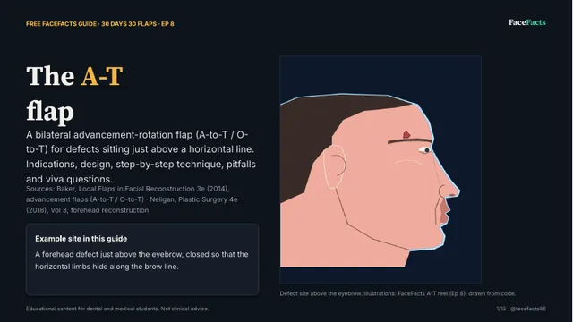
30 Days 30 Flaps, Ep 8: the A-T flap (PDF on Google Drive, opens in a new tab)
A bilateral advancement-rotation flap for defects just above a horizontal line: indications, design, steps, pitfalls and viva questions.
Cited PDF presentations that go with our Reels and carousels. Free to read and keep, no sign-up. Each one opens in Google Drive.

A bilateral advancement-rotation flap for defects just above a horizontal line: indications, design, steps, pitfalls and viva questions.

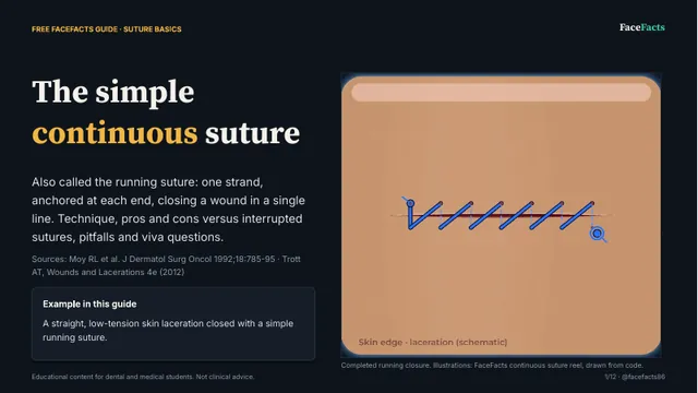
One strand anchored at each end: technique, pros and cons against interrupted sutures, pitfalls and viva questions.
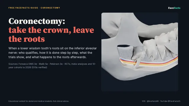
When wisdom tooth roots sit on the IAN: who qualifies, the technique step by step, what the trials show and what happens to the roots.

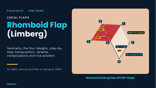
60°/120° geometry, the four designs, step-by-step transposition, variants, complications and viva answers.

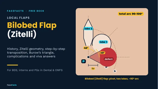
History, Zitelli geometry, step-by-step transposition, Burow's triangle, complications and viva answers.

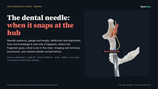
Gauge, deflection and aspiration, why breakage happens, where the fragment goes, what to do in the chair, retrieval and prevention.

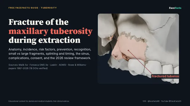
Anatomy, risk factors, prevention, small vs large fragments, splinting and timing, the sinus, complications and consent.

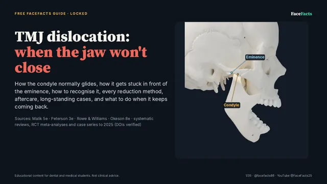
How the condyle gets stuck in front of the eminence, recognising it, every reduction method, aftercare and recurrent cases.

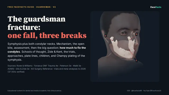
Symphysis plus both condylar necks: mechanism, the open bite, how much to fix the condyles, approaches, plate lines and children.

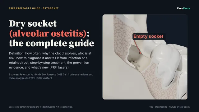
Why the clot dissolves, who is at risk, telling it from infection, step-by-step treatment and the prevention evidence.

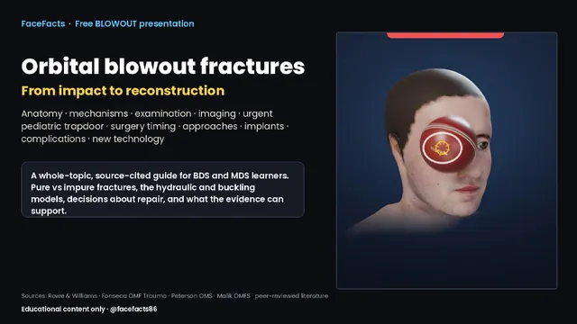
Why the rim holds and the floor gives way: signs, forced duction, CT, the paediatric trapdoor and repair materials.

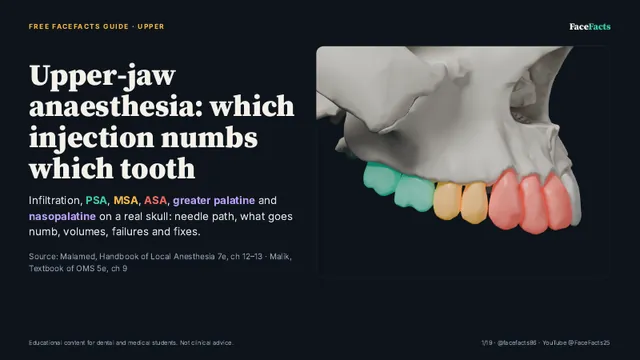
Infiltration, PSA, MSA, ASA, greater palatine and nasopalatine on a real skull: needle paths, volumes and what each one numbs.

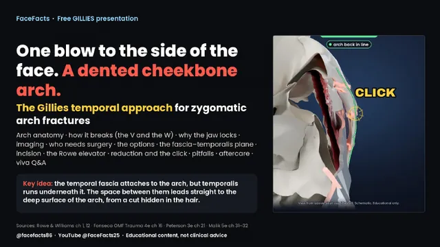
How the arch breaks, why the jaw locks, and the temporal approach that lifts it back through a cut hidden in the hair.

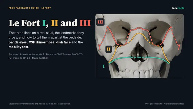
The three lines on a real skull and how to tell them apart at the bedside: panda eyes, CSF rhinorrhoea, dish face, mobility.

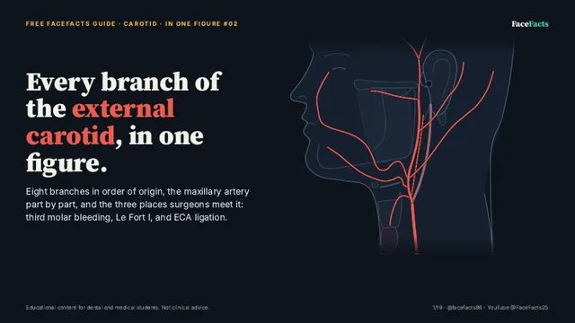
Eight branches in order of origin, the maxillary artery part by part, and where surgeons meet them.

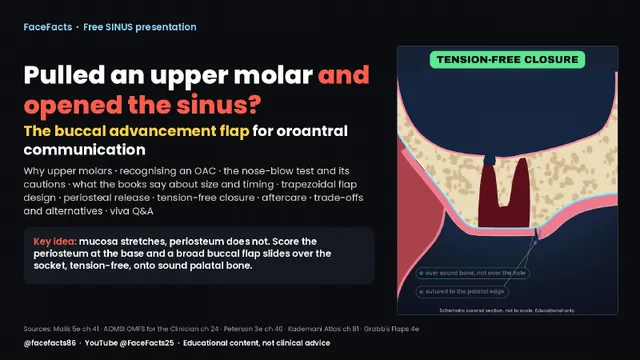
Recognising an OAC, the nose-blow test and its cautions, size and timing, and a tension-free flap closure.

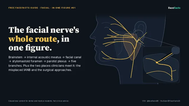
Brainstem to the five branches in one figure, plus the misplaced IANB and the surgical danger zones.

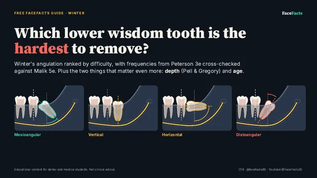
Winter's angulation ranked by difficulty, plus the two factors that matter even more: depth and age.

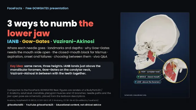
IANB, Gow-Gates and Vazirani–Akinosi: targets, landmarks, depths and when to choose each.

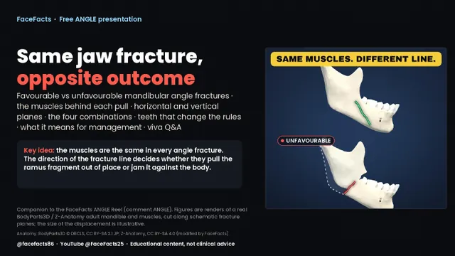
Same muscles, different line: how the direction of the fracture decides whether the fragments stay put.

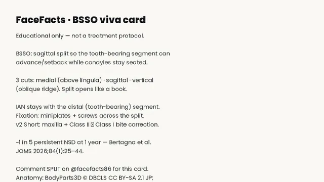
The bilateral sagittal split osteotomy on one page: the three cuts, the split, fixation and the nerve.

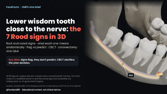
The seven root and canal signs in 3D, what each means anatomically, flag vs predict, CBCT and coronectomy.

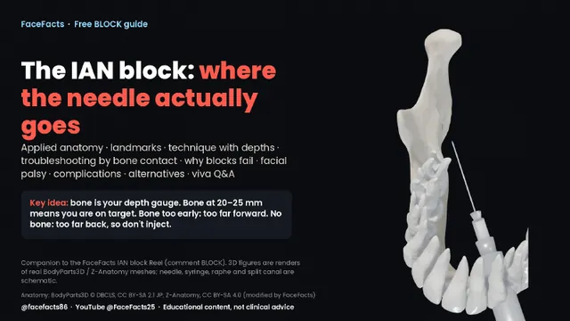
Applied anatomy, landmarks and depths, troubleshooting by bone contact, and why blocks fail.

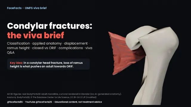
Classification, applied anatomy, displacement, ramus height, closed vs open treatment and complications.
On Instagram? Comment the keyword on the matching post (for example UPPER) and we'll send you the same PDF by DM.
Spotted an error? Message us on Instagram @facefacts86. Corrections are welcome and get fixed.